CLS User Office
The CLS User Office is available to help navigate CLS user portal and other related information. When submitting BTR, proposals, etc. the USO will communicate with the user the necessary trainings, etc. up to 6 weeks prior to the schedule beamtime. For more informationcontact the USO-Support
Contacting Beamline Staff
Beamline staff are available to help provide a smooth beamtime for the users. When not on site, the beamline staff use Skype on the windows machine where users are able to communicate with the beamline staff.
Sample Storage
If you arrive with samples that require storage in either Ar or N2 ( Rm 1080) Glovebox. Please refer to the following page for future assistance or contact our CLS Lab Manager Burke Barlow
Setting up a XAS Scan
Setting up a XAS Scan
- How do I setup a XAS Scan?
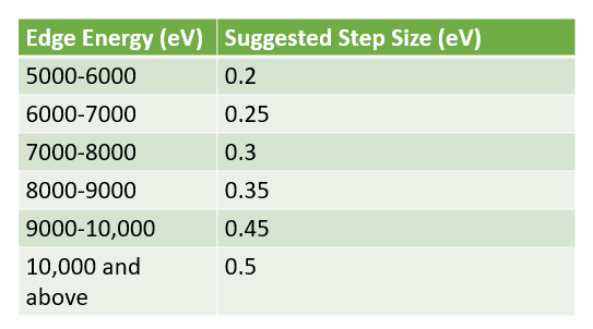
- Accquaman does a good job in predetermining the regions for XANES and EXAFS. Generally as energy is scanned across, the absorption cross section decreases, and EXAFS signal must be improved by longer count rates or by averaging across several scans. For XANES, the limitations in the step size (vs. oversampling) is the resolution of the crystal set used. Generally, a guide below can help provide information for setting up a step size in the XANES region. For EXAFS a typical spacing of of 0.05 inverse angstrom ( ~ 0.2E1/2) is reasonable.
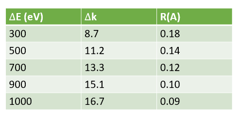
- How far out in X(k) can I measure?
- The EXAFS oscillations die as 1/E. S/N is proportional to sqrt of the # of scans. Careful interplay of statistic and time should be taken into account.
- Generally, one can collect 1000eV above the edge, the uncertainty in will plateau, and are only able to radially resolve at most a difference of 0.09A
- I have a sample that I am measuring in transmission mode, and I don't see the reference signal? What does this mean?
- This would mean that your sample is too thick, and is absorbing and reducing the amount of transmitted beam to the IC for the foil. The advice would be to reduce sample thickness
- I have a sample that is supported sample, I know that it has adequate composition of the element of interest in transmission mode, but I don't see a good signal in the Foil IC or in transmission.
- This would mean that your support even at this energy is absorbing some of the beam, and reducing the beam going into the foil IC. Reduced sample thickness or number of tapes.
- There a systematic glitches in my transmission data - what is it?
- It can either be due to glitches in I0 ( check the signal in I0) or it can be what is referred to as a pinhole effect, where the beam sees no sample on the beam scale or it can be that the crystal size are too large that there is signal sample diffraction
Collecting Data Using Accquaman
When I started my measurements, the shutters closed?
- This is normal, Accquaman measures dark currents before the start of the scan. The dark current scan takes as long as the maximum dwell time used in the region file for the XAS scan. For example, if you measure EXAFS up to 14 k, and the scan has a 10 s dwell time, then the dark current measurements will take 10 s to collect. If measuring XANES with a 1 s/point dwell time, then dark currents will measure for 1 s. In some occasions, the Phoebus panel update, the indicator for the photon shutter maybe be closed for longer, but the ACIS panel will automatically update in real time. As a precaution, look at the ACIS panel to ensure that after dark current measurements, the photon shutter is open
Why is Acquaman lagging?
- It is advisable to restart Acquaman program every day. This will hep reduced the software lag.
I am having trouble with energy calibration using a standard foil. When I hit the calibrate button, nothing seems to happen.
- Yes, this is normal, only click the calibrate button once and that will allow the software to more to the desired position. If the calibrate button is clicked more than once, it will shift the energy the same magnitude as the shift between the absolute energy and calibrant foil energy. Please only click once.
What is the correct way to calibrate?
- Generally, use the first derivative peak. See Reference X-ray Spectra of Metal Foils
My scan is running but the acquisition window stopped showing the spectra being collected. What should I do?
- It is likely that there is a change in the scale that you are viewing. Re-adjust the scale and you should see your scan.
I have low counts on my reference spectra when doing the calibration, why is that?
- Make sure that the reference foil on the reference foil wheel is properly aligned.
I am on my sample and ready to measure, but I don't see any counts on the Ge detector.
- Make sure that the photon shutter is open, the Ge-detectors are initialized and that the caps are removed.
I have calibrated and I am ready to find my sample in the beam. I tried to find the sample but I could not, what do I do?
- Use the fluorescent screen to verify that the beam is on your sample. If you see the beam on sample, then the sample concentration for the element of interest is low, or the sample is too thick ( absorbing)-you can check if you are getting signal from your reference ionization chamber, or the sample is too thin and the amplifier is saturated ( i.e. the voltage reading is above 5V). If you have a standard sample with known concentration it is advisable to measure the standard samples to see if you are getting expected signal and signal quality.
Acquaman just crashed and the software closed abruptly- How do I troubleshoot?
- Navigate to desktop screen and restart Acquaman.
I restarted Acquaman but now I don't have counts in my ICs
- Navigate to Scaler, change the counts from single shot to continuous.
There is a huge spike ( downward spike) when measuring that is more intense than the counts/signal from the XAS data I collected, and I see it only in the Dark Corrected Spectra? What is this?
- It can be a few things ( ice diffraction, contamination of a different edge, or incorrect dark current counts).
- If ice diffraction, and measuring using Ge detector, not all pixels will capture the diffraction, and the signal will be more localized ( i.e. some pixels will see it, other pixels will not see it). Check each channel,
- If contamination of different edge, corroborate this with the edge finder on Hephaestus ( typically for Cu, there is almost a Zn signal),
- and finally if in correct dark current, the spike will only be present in the dark corrected spectra, and you are probably getting negative counts because CSS was measuring dark currents with the beam accidently turned on- shutters were open (Acquaman automatically shuts the beam during this process), please make sure beam is off for dark current measurements). Change the gains, and the offsets will be adjusted accordingly.
I stopped the scan because I need to measure with Ge detector instead of PIPS. How can I restart collecting Fluorescence with the Ge detector?
- In the Data Collection Tab in Acquaman, make sure to tick the channels for In and Outboard Ge detector. If switching back to PIPS, navigate to configuration tab in Acquaman and click on PIPS to configure for PIPS.
Why are shutters not closing in the beginning of the scan?
- It maybe an Acquaman bug, please make sure to restart Acquaman and make sure only one instance is open The shutters close at the beginning of the scan to measure dark current. You will see that the POE shutter will be RED and turn GREEN after dark measurements are collected.
I am using the Ge detector to measure my sample, but I do not see any counts when running test scan?
- If you are on the sample, with correct position and beam slit size, make sure that the correct fluorescence line for the elements of interest is selected, otherwise it will read out no counts.
In the event of a beam dump
- Check Machine Status or login in to the user portal to follow updates on machine status.
- In the event of a trip, ring dump, etc., the machine status will change. With no beam, you can choose to pause or cancel your scans and wait for the machine status to show that the ring current is ~220 mA and that the ring is in top up mode.
- How long do I need to wait for the optics to warm up after beam-dump?
- If it was a abrupt dump which was recovered quickly. You may wait the duration of the dump for warm-up. Generally, rule of thumb, wait the duration of the beam-dump for warm- up but no more than 2 hours.
Cryocooler and Cryostat
- After a sample swap, the cryostat temperature started to decrease slowly, we tried to flush and opened the needle valve a little bit, but there was no detectable temperature change. How can we get the cryostat to cool down to 80K faster?
- First it is important to check that the needle valve on the cooling pump is open. The needle valve is fully open after 6 full rotations. In order for adequate cooling, 2-3 turns is sufficient. Please ensure you have enough LHe or LN2 when doing so. Monitor the cryo-insulation pressure, the reading should be around 6-5 E-5 torr. Please be careful when opening the needle valve and if uncertain if the valve is closed or open. Fully close the valve and then take note of the number of rotations.
- My sample fell in the cryostat, what should I do?
- It may be a good idea to warm the cryostat to 300 K, disconnect the lines and flip the cryostat under a fume hood.
Detectors
- I am using the Ge detector, what is an acceptable count value?
- Each of the green windows on the Ge detector software represent a pixel. The Ge detector we have has 32 pixels. Each pixel count rate should not exceed 150 ( i.e. 150k counts).
- What does a filter do?
- A filter will absorb the scattered peak, while transmitting most of the fluorescent line of interest. A filter can improve the S/N. For more information visit X‐ray filter assembly for fluorescence measurements of x‐ ray absorption fine structure.